 Therapy by Alice
Treating Neck Cricks From the Eric Dalton Blog
Posted on May 1, 2020 Leave a Comment
Researchers do not know if impaired muscle function is the primary cause of joint dysfunction, or if the reverse holds true. However, we do know that a reflexogenic relationship exists between muscles, joints and the nervous system. As components of the spinal anatomy begin to degrade over time, bone loss, disc degeneration, and facet joint osteoarthritis may place excessive stress on the aging vertebrae. The body responds by growing bony nodules called osteophytes, or bone spurs, to compensate for diminished spinal stability.

While the word spur often leads people to imagine something sharp or pointed digging into a nerve or other tissue, bone spurs are actually smooth and sometimes crusty growths that can often be mobilized through movement. Although bony spurs are common and do not exhibit symptoms in most cases, when they grow in confined areas adjacent to nerve roots or the spinal cord, nerve compression can occur. Because the uncinate processes are located near the foramina – channels where nerve roots exit the spinal canal – bone spurs that may develop at the uncovertebral joints resulting in a condition known as foraminal stenosis (Fig. 1). Should this narrowing of the foraminal canal lead to nerve compression, the brain may deliver symptoms such as localized or radiating pain, tingling, numbness or protective guarding.
So, what can manual and movement therapists do to treat neck cricks caused by facet jamming and nerve root impingement? In many cases, protective muscle spasm can be reduced through application of specific soft-tissue decompression and mobilization maneuvers that help restore “joint play” and facilitate hydration of flattened intervertebral discs. In the video clip seen here, I demonstrate three basic, but effective, myoskeletal techniques to relieve “immobilization arthritis” due to facet jamming and bone spur formation.
Treating Neck Cricks
GOAL: Mobilize cervical muscles that are restraining joint motion
LANDMARK: Typical vertebrae (C2-3 to C7-T1)

A. ACTION: Chin Jutting (client supine)
- Therapist’s hands come together so his thumbs can create a fulcrum to brace on both sides of the client’s spinous processes in the lamina groove
- Therapist brings thumbs ½ inch apart and places them into the lamina groove anywhere in the mid-cervical spine
- Therapist gently mobilizes client’s neck by pushing up with both thumbs into the groove
- This rhythmic action juts the client’s chin as each vertebral segment is extended over the thumb fulcrum
- Repeat the chin-jutting maneuver for 2 minutes and reassess for increased mobility

B. ACTION: Cervical sidebending to relieve nerve root compression
- Therapist’s cupped palms support client’s head as both thumbs pin the lateral body of the C5 spinous process
- Therapist’s hands right sidebend client’s neck while the right thumb presses against the side of the spinous process (This creates a fulcrum at 5 that tests the mobility of the joint capsules)
- As therapist steps to his right side he creates a fulcrum with his right thumb
- Therapist repeats by sidebending left and pushing with the left thumbIf a right sidebending restriction is found as the therapist examines up and down the cervical spine, client inhales and gently left sidebends head against therapist’s resistance to a account of 5 and relaxes
- Therapist right sidebends client’s neck to new restrictive barrierRepeat this maneuver until a release is felt in the fibrotic capsules and lamina groove muscles

C. ACTION: Translation/ Undulation for nerve root mobilization
- Therapist’s thumbs face inferiorly and brace each side of the client’s spinous processes
- As therapist steps to his left foot, his right thumb pushes against the spinous processes
- As he steps to his right foot, his left thumb presses against the spinous processes
- Therapist begins a rhythmic undulating motion as he rocks back and forth foot-to-foot, pushing on bodies of spinous processes
- Therapist examines for motion restricted tissues from C2-3 to C7-T1 and reassesses for improved mobility
An Easy and Healthy Recipe
Posted on April 29, 2020 Leave a Comment
https://www.abmp.com/updates/blog-posts/five-easy-and-healthy-recipes
Now is an ideal time to explore cooking new recipes while we’re all encouraged to stay home during the pandemic. These five recipes from ASCP Skin Deep magazine contain healthy ingredients like antioxidant-rich vegetables and vitamin-rich fruits. In case you need new ideas to shake up your routine, you can make these delicious recipes for lunch, dinner, or even a quick snack. These recipes are all vegetarian. The dark chocolate chip cookies are flour- and refined sugar-free, and the yellow curry bowls are gluten-, dairy-, and sugar-free. Also, the ginger pear smoothie and the kale, peach, and cabbage salad are both gluten-free. Bon appetit!
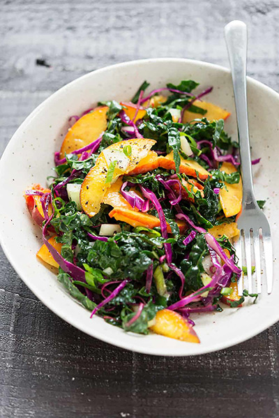
Try this collagen-boosting triple threat containing vitamin C-rich kale, peaches, and red cabbage.
1 bunch kale, preferably dinosaur/lacinato
1 cup finely shredded red cabbage
1 cup cucumber, peeled, seeded and thinly sliced
2 scallions, thinly sliced
2 tablespoons extra-virgin olive oil
2 tablespoons fresh lime juice
1/2 teaspoon salt
1/4 teaspoon freshly ground black pepper
2 ripe peaches, thinly sliced
In a large bowl, toss together the kale, cabbage, cucumber, and scallions.
Drizzle on the olive oil, lime juice, salt, and pepper, then massage into the slaw for 1–2 minutes to gently wilt the kale and the cabbage. Taste, adding more salt, pepper, lime, or olive oil as needed.
Add the peaches and toss again.
Read more by clicking the above link or stayed tuned for more…
BOGO Flash Sale
Posted on April 29, 2020 Leave a Comment

Flash Sale!!!! Buy one get one. It’s a great time to stock up. Message me for more details, or drop a comment below.
Another Healthy Recipe
Posted on April 29, 2020 Leave a Comment
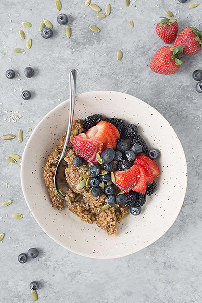
Super Seedy Baked Oatmeal
This baked oatmeal is rich in gamma-linolenic acid, or GLA, an essential omega-6 fatty acid that helps your body lock in moisture for healthier, more supple skin.
- 2 tablespoons ground flax seeds
- 1/4 cup water
- 1 large ripe banana, sliced
- 1 1/2 cups quick cooking oats
- 1/4 cup packed brown sugar
- 1/2 teaspoon cinnamon
- 1/4 teaspoon ground ginger
- 1/8 teaspoon ground nutmeg
- 1 teaspoon baking powder
- 1/4 teaspoon salt
- 1/2 cup + 2 tablespoons hemp milk (or other nondairy milk)
- 1/3 cup pure maple syrup
- 1 teaspoon vanilla extract
- 2 tablespoons melted coconut oil
- 1/4 cup pepitas (shelled pumpkin seeds)
- 2 tablespoons hemp seeds
- fresh berries, for serving
Preheat oven to 350 degrees F. Lightly spray an 8-inch square pan with nonstick spray.
Whisk together the ground flax seeds and water in a small bowl, then set aside to gel.
Slice the banana and place in a single layer on the bottom of the square pan.
In a separate bowl, mix together the oats, brown sugar, cinnamon, ginger, nutmeg, baking powder, and salt. Add the flax seed mixture, hemp milk, maple syrup, vanilla, and coconut oil. Stir together, then fold in the pepitas and hemp seeds.
Gently top the sliced bananas with the oat mixture. Place in the preheated oven and bake for 30 minutes until golden brown and set.
To serve, scoop the baked oatmeal into a bowl and top with fresh berries and more hemp milk.
Robert Gardner Podcast
Posted on April 28, 2020 Leave a Comment
www.instagram.com/tv/B_UG1CQHjET/
I have been learning so much from Robert. I can hardly wait till I can finally get to his Thai Yoga jam in Austin.
Heart and Harmony Music Therapy
Posted on April 26, 2020 Leave a Comment
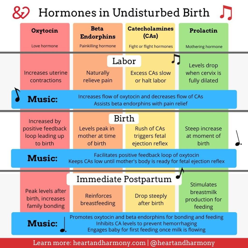
Nikki Belshe is a brilliant music therapist and she is just killing it. I wish I had as much creativity in my pinky as she has in her entire body! She is already doing the work I would like to be doing. Thanks Nikki for sharing this valuable too.
Understanding Covid 19’s Cytokine Storm
Posted on April 22, 2020 Leave a Comment
Although our understanding of how the coronavirus affects the body is still very incomplete (and changing rapidly), we do know the virus’s effects vary tremendously from person to person. Some infected people have no symptoms at all; others have a cough and sore throat; some need medical care and may even die as their inflamed lungs and organs fail. Evidence is mounting that in a number of those fatal cases, it isn’t the virus itself that kills the host, but rather, it is the body’s own out-of-control inflammatory reactions, snowballing into a “cytokine storm” that deals the fatal blow.
So, what are cytokines and how can they kill? Does this help us understand anything about other kinds of inflammation? WHAT ARE CYTOKINES?
When the body’s immune cells detect pathogens or tissue damage, they secrete cytokines (Image 1) to signal, mobilize, and instruct other immune processes and cells. The word cytokine, in fact, comes from the Greek κύτος (kytos) or “cell,” plus κίνησις (kinēsis) or “movement.” Simplified, cytokines are cell movers. Though cytokines are relatively simple protein molecules, their complex functioning and interactions are still being deciphered. We know cytokines can be inflammatory (turning up immune processes); anti-inflammatory (turning off inflammation); or inflammation-resolving
(by signaling “next” in the progression of normally self-resolving inflammatory phases that lead toward healing). In other words, cytokines orchestrate, modulate, and time each phase of normal inflammatory progression. Except in some cases, this orchestration goes very wrong. When a virus like COVID-19 infects the lungs, it hijacks cells there and turns them into virus factories. This damages the cells and releases inflammatory cytokines. These acute-phase cytokines have several important effects:
• They painfully irritate nearby nerve endings, triggering the aches and pains typical of influenza (causing the host to modify its behavior, and rest).
• They summon other immune cells (to attack the invader and damaged tissues). Cytokines are the immune system’s inflammatory messengers and coordinators: when immune cells detect pathogens or tissue damage, they secrete cytokines to signal and regulate inflammatory cells and processes.
Although our understanding of how the coronavirus affects the body is still very incomplete (and changing rapidly), we do know the virus’s effects vary tremendously from person to person. Some infected people have no symptoms at all; others have a cough and sore throat; some need medical care and may even die as their inflamed lungs and organs fail. Evidence is mounting that in a number of those fatal cases, it isn’t the virus itself that kills the host, but rather, it is the body’s own out-of-control inflammatory reactions, snowballing into a “cytokine storm” that deals the fatal blow.
• They make cell and vessel walls leaky (which helps transport the important immune cells and chemicals to where they’re needed most).
So far, so good—all of these inflammatory functions are healthy, normal, and needed. In most cases, the immune
system’s anti-inflammatory and resolution-
triggering mechanisms come to bear, winding down these acute inflammatory reactions. Tissue healing progresses, sensation normalizes, and function returns. THE CYTOKINE STORM
But, for reasons we don’t entirely understand, in about 15 percent of people battling any serious infection,1 the immune
cycle seems to get stuck in a raging release of more and more cytokines, leading to more cell death, and even more cytokines. Perhaps related to underlying genetic defects,2 this phenomenon (first described in
1993) has been long studied in related forms such as systemic inflammatory response syndrome, cytokine release syndrome,
macrophage activation syndrome, hemophagocytic lymphohistiocytosis, or cytokine storm syndrome (CSS).3 In the
1918 Spanish flu epidemic, differences in cytokine storm reactions between old and young, and between that virus and COVID-19, are thought to explain why that epidemic killed so many young people. CSS doesn’t just occur in influenza, but in other respiratory diseases caused by coronaviruses, as well, such as SARS and MERS. They are also associated with noninfectious diseases such as multiple sclerosis and pancreatitis. But whenever cytokine orchestration has turned into
a cytokine storm, their normally helpful
functions contribute to an escalating spiral of worsening symptoms:
• The pain, sensitivity, and fatigue from influenza’s cytokines can make the host inhibit breathing and movement, so much that the concentrated and by-
now toxic inflammatory soup collects in the lungs’ alveoli (Image 2).
• The cytokine-summoned first-responder cells are aggressive and indiscriminate in their destruction. Flooding into the tissues on the wash of leaked interstitial fluids, they use powerful enzymatic and oxidative processes to demolish both friend and foe alike (Image 3). Other cells, such as fibroblasts, simultaneously try to repair this ongoing damage, but when the cytokine-induced destruction continues, New! ABMP Pocket Pathology at http://www.abmp.com/abmp-pocket-pathology-app. 81
the resulting excess collagen makes tissues denser and more fibrous, further inhibiting normal perfusion and drainage.4
• In musculoskeletal tissues, the plasma leakage of acute inflammation causes its characteristic swelling and redness. In lung tissue, this fluid buildup causes coughing; and if excessive or prolonged, can lead to breathing difficulties and pneumonia. What’s more, capillaries damaged by the inflammatory riot can let the cytokine-rich fluids spill over into the bloodstream, resulting in systemic inflammation and multi-organ failure.5
Microdamage to the lungs’ alveoli from virus replication can, in some cases, trigger a self-escalating “cytokine storm” of hyperinflammatory cytokine overproduction and further tissue damage.
WHERE DOES THIS LEAVE US?
If there’s good news in the cytokine storm story, it might be that these cytokine imbalances appear to be detectable (and to some extent, treatable) in COVID-19 patients via commonly available blood tests and immunosuppressive drugs. When excessive inflammation is found in a coronavirus patient, doctors face a tricky decision about how much to suppress the immune system in the face of an active infection, but as experience dealing with this virus mounts, protocols will emerge to help guide these treatment decisions. As has happened with past epidemics, the flood of attention, awareness, and resources will increase our overall understanding of how inflammation gets out of control in other conditions. And there are many, many of those conditions: virtually all musculoskeletal complaints, the majority of chronic diseases, and a growing list of psychological and behavioral conditions are now understood to have a primary inflammatory component. For now, many bodyworkers, manual therapists, and massage therapists are restlessly watching the COVID-19 story unfold from the sidelines. We’re looking and learning while in this holding pattern, but at some point, we’ll have our work to do: helping people stay healthy now has even more important implications than it used to. Immune competence is emerging as a key factor in overall health, and we have clear contributions to make there. As time passes, we will learn more about how we can support post-coronavirus recovery of both individual survivors, and of our world.
Notes
1 Cytokines are the immune system’s inflammatory messengers and coordinators: when immune cells detect pathogens or tissue damage, they secrete cytokines to signal and regulate inflammatory cells and processes.
2 Microdamage to the lungs’ alveoli from virus replication can, in some cases, trigger a self-escalating “cytokine storm” of hyperinflammatory
cytokine overproduction and further tissue damage.
3 Lung tissue sample from a 1918 influenza victim, showing extensive damage to alveoli from inflammatory cell infiltration.
- R. Cron, as quoted in Apoorva Mandavilli, “The
Coronavirus Patients Betrayed by Their Own
Immune Systems,” New York Times, April 1,
2020, http://www.nytimes.com/2020/04/01/health/
coronavirus-cytokine-storm-immune-system.html. - Grant S. Schulert et al., “Whole-Exome Sequencing
Reveals Mutations in Genes Linked to Hemophagocytic
Lymphohistiocytosis and Macrophage Activation Syndrome in Fatal Cases of H1N1 Influenza,” Journal
of Infectious Diseases 213, no. 7 (April 2016):
1180–88, https://doi.org/10.1093/infdis/jiv550. - J. L. Ferrara, S. Abhyankar, and D. G. Gilliland,
“Cytokine Storm of Graft-Versus-Host Disease: A
Critical Effector Role for Interleukin-1,” Transplantation Proceedings 25 (February 1993): 1,216 –17. - J. R. Tisoncik et al., “Into the Eye of the
Cytokine Storm,” Microbiology and Molecular Biology Reviews 76, no. 1 (2012): 16–32,
https://doi.org/10.1128/MMBR.05015-11. - Fields Virology, 6th ed., eds. David M. Knipe and
Peter M. Howley (Philadelphia: Wolters Kluwer/
Lippincott Williams & Wilkins Health, 2013).
Til Luchau is the author of Advanced Myofascial Techniques (Handspring Publishing, 2016), a Certified Advanced Rolfer, and a member of the Advanced-Trainings.com faculty, which offers online learning and in-person seminars throughout the United States and abroad. He invites questions or comments via info@advanced-trainings.com and Advanced-Trainings.com’s Facebook page.
Coronavirus…Again
Posted on April 15, 2020 Leave a Comment
Coronavirus got me like…. 🦠
- Thankful I work from home and not with little kids everyday. 😷
- Yes I’m out and about living life but am taking our immunity, and super reds and super greens like crazy 🙃
- I’m getting to ride my horses more often than if I was working and I haven’t had to worry about paying the bills as much as if I hadn’t had this business and my health.
Y’all I have the ✌🏻 solutions for you 😜. Come join me and make money from your home + I got the best IMMUNITY BOOSTERS ever.
Coronavirus ain’t gonna be touching me ✌🏻 We are all in this together


